Speed Meibography

Speed meibography. Red free, Monochrome, and Angiography
For all DED exams
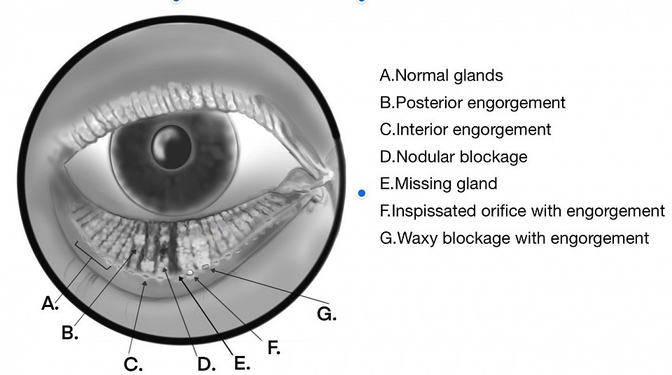
The new Speed Meibography LED is a significant advancement in DED consultations. It examines both the oil glands and accessory lacrimal glands. Meibography can be done on every slit lamp. By simply clipping on the light tower the special red-free LED renders the glands. You can correlate DED clinical signs and symptoms. The ability to assess not only gland loss but also blockage and engorgement provides valuable insights into gland function. An added benefit is visualization of the accessory lacrimal glands located behind the tarsal plate. Overall, Meibography's value in diagnosing oil film abnormalities and structural gland issues is indispensable for addressing dry eye patients effectively.
Top is red free green meibography
Middle Black and white using camera
Bottom Inverse to show angiogram of glands
Visualize Acinar Structures
Incredible detail can be found by increasing magnification on your slit lamp. Unlike infrared meibography the detail of each acinar structure can be seen. The only limitation is optical zoom’s resolution. This is important since you want to discover problems before the gland has perished. The acinar spaces make meibum allowing us to correlate structure to function.

Comparison of histology to speed meibography
Meibography
Speed meibography can be done in red free green or black and white using a camera. Either shows abnormalities. The diagram demonstrates the common abnormalities. This adds to the traditional meiboscores by being higher in resolution and showing the acinar structures.
Acinar meibography

Monochrome conversion from red free illumination
Use of the EyePhotoDoc camera to analyze and track treatment
The camera along with Apps allow you to document findings. You can even make them into black and white monochromes to resemble the IR meibography. Indeed that will increase contrast and make it feel ‘familiar’.
Using the finger stretching of the photo allows you to enlarge the area of interest for easier diagnosis.
Nodular changes
Missing gland
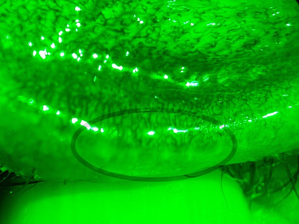
Orifice obstructions
Bloated glands

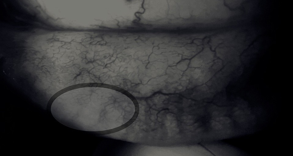
Angiography showing gland loss

High power shows missing gland
Case study
When you study this low magnification photo can you see the abnormalities of the glands? Using your slit lamp you can zoom up and see incredible detail. Remember your eyes have 40 times the resolution of the 12 megapixel camera!
Find:
1. Missing glands
2. Dilated acinar structures
3. Missing acinar structures in gland
4. Segmented glands
5. Fun bonus question Can you see the accessory lacrimal glands?
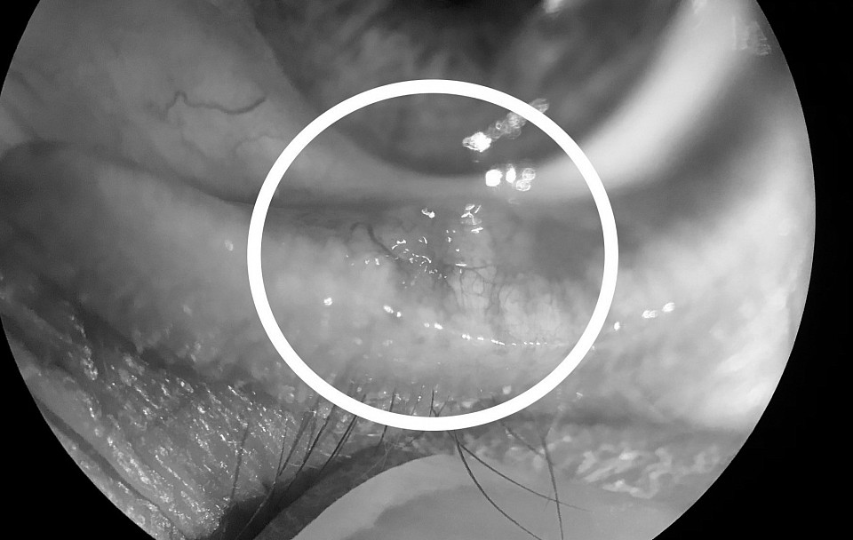
Can you find the abnormality?

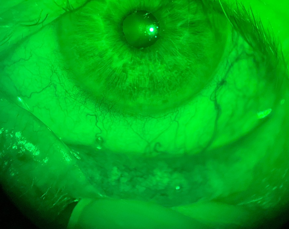
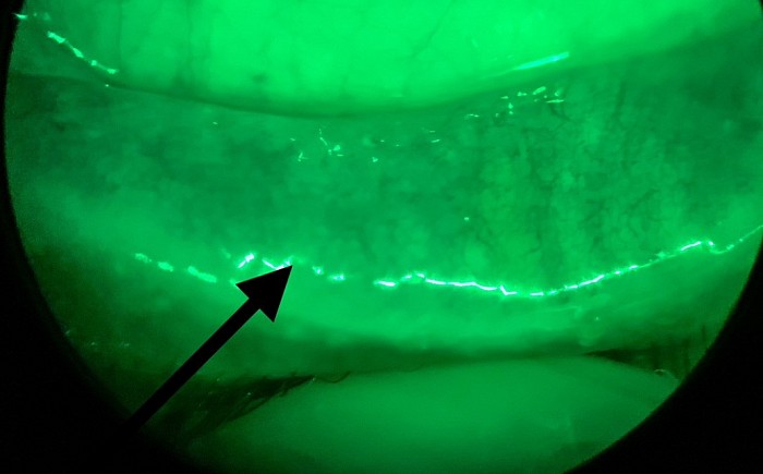
Precorneal tear film
Clinical diagnosis MGD
The meiboscores are an overview based on gland’s central tubes. We have evolved that evaluation based on the ability to see acinar structures and vasculature.
1. Look for loss of multiple glands especially in an area which causes symptoms and the signs offluorescein stain on the cornea or lissamine green stain on the conjunctiva.
2. Examine if the acinar spaces bulge out representing impaction or oppositely focal reduction of acinar structures.
3. View for tortuosity of glands
4. Reduced blood vessels over devitalized meibomian structures
5. Function test If a gland abnormality is present have patient squeeze the lids firmly to see if normal particles are released into the tear film.
6. Meibomian expression Note ease of expression guided by findings. (Meibomian is made up of not only oils but also waxes and proteins. Current research finds the ratio of oil decreases to waxes or proteins and causes expression difficulties). Oils are liquid at room temperature but waxes and proteins are solids.
7. Examination of the precorneal tear film for microsphericles and debris.
